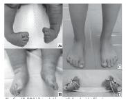
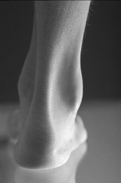
OverviewPes cavus is defined as foot having an abnormally high medial longitudinal arch.. These feet retain their high-arched appearance when weight bearing, this is the supinated foot type. It What is the tendon at the back of your ankle? a less common deformity than flat foot (pes planus). Pes cavus is usually bilateral and apparent at an early age. The sudden appearance of the deformity, or its presence unilaterally, may be the result of trauma or neuro-muscular disease.
CausesMost cases of high arches are associated with nervous-system disorders. The conditions that can cause high arches include Cerebral palsy, Spina bifida, Muscular dystrophy, Polio, Stroke, Charcot-Marie-Tooth disease, Spinal cord tumor. The cause of high arches cannot be determined in about one in five instances. These cases are called idiopathic, meaning the condition arises from an unknown or uncertain cause.
 Symptoms
Difficulty finding proper fitting footwear because the shoes are not deep enough due to high arch and the clawed toes. Shortened foot length. Foot pain with walking, standing, and running. Metatarsalgia with pain in the forefoot/ ball of the foot (usually 1st and 5th metatarsal heads), with or without calluses/corns. Pain and stiffness of the medial arch or anywhere along the mid-portion of the foot. Morton's neuroma with pain in the ball of the foot and lesser toes. Pain in the heel and sole of the foot from plantar fasciitis. Stress fractures of the metatarsals and other foot bones. Particularly in diabetics and those with compromised circulation, abnormal pressure may result in chromic ulcers of the heel and ball of the foot. Strain and early degenerative joint disease (osteoarthritis) of lower extremity joints. ?Pump bumps" (Haglund's deformity) on the back of the heel. Associated discomfort within and near the ankle joint. Ankle instability with frequent sprains. Tight Achilles tendons. The knees, hips, and lower back may be the primary source of discomfort. Chronic lower extremity pain my lead to inactivity and diminished well-being.
DiagnosisExamination of the muscle groups and muscle strength is important. Furthermore, pain along the peroneal tendons may be a sign of a peroneal tendon tear. This may result in a cavus foot much like a posterior tibial tendon dysfunction may result in flatfoot. Instability of the lateral ankle may also lead to a cavus foot position as the talus deviates into a varus position due to the laxity of the lateral ankle ligaments.
Non Surgical TreatmentConservative care is highly successful in the cavus high arch foot. An orthotic with a high lateral heel flange, a valgus post and a sub-first metatarsal cutout can balance the foot. Often, the first ray is plantarflexed and a cutout of the first metatarsal head is essential for forefoot balancing. In severe ankle instability cases, an over the counter ankle-foot orthotic or a custom ankle-foot orthotic can be beneficial in balancing the foot and ankle. Consideration of a first ray cutout should also be part of the bracing process.
Surgical TreatmentIf the above techniques do not help relieve pain and provide stability, two surgeries within the span of two weeks may be needed. During the first surgery, the orthopedic surgeon releases the tightest of the soft tissues in the arch, which causes the rest of the tissues to relax. During the second operation, the surgeon uses a bone graft to reshape at least one bone and moves several tendons to improve muscle balance.